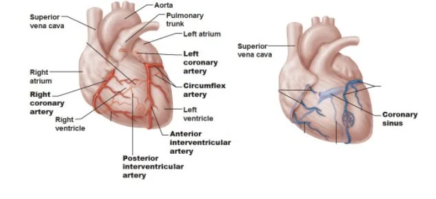
Blood Supply to the Walls of the Heart
Similar to the rest of the body, oxygenated blood from the heart is supplied to itself to ensure it is supplied with the nutrients and oxygen it needs to maintain its function.
Right coronary artery
It supplies the right atrium and ventricle. Its origin is above the anterior flap of the aortic valve. It runs forwards between the right auricle and pulmonary trunk, then descends in the right atrioventricular groove and continues on to the base of the heart along the groove where it anastomoses with the left coronary artery.
It’s marginal artery is a major branch and it runs along the right border of the heart towards the apex.
It’s posterior interventricular artery is it’s second major branch and it runs in the posterior interventricular groove towards the apex.
Left coronary artery
It originates above the left flap of the aortic valve. It passes behind and to the left of the pulmonary trunk and follows the left atrioventricular groove until it anastomoses with the right coronary artery on the base of the heart.
It’s anterior interventricular artery branch arises at the anterior interventricular groove and follows it to the apex. The anterior interventricular artery usually ascends a short distance in the posterior interventricular groove to anastomose with the posterior interventricular artery. The anterior and posterior interventricular branches supply blood to both ventricles.
It also has a left marginal branch running down the left border of the heart to the apex.
Potential anastomoses exist between the various branches of the coronary arteries where the meet on the base of the heart and the apex. These are, however, inadequate to bypass a sudden blockage of a large artery.
Venous Drainage
Most of the coronary veins accompany their corresponding coronary arteries. The veins that drain the heart join to form the coronary sinus in the posterior atrioventricular groove. About two-thirds of the venous blood from the heart wall drains into the right atrium by this route.
The tributaries of the coronary sinus include:
Great cardiac vein
Middle cardiac vein
Small cardiac vein
The anterior cardiac vein, draining most of the anterior surface of the heart, empties directly into the right atrium. The remainder of venous blood drains by small veins in the chamber walls that empty directly into the heart chambers.
Clinical Notes:
Myocardial infarction occurs as a result of a blockage of coronary vessels.
Angina pectoris occurs from ischemia and less severe blockage of coronary vessels.
Coronary artery bypass graft utilizes the great saphenous vein or radial artery.
Coronary angioplasty utilizes a small inflatable balloon and stent to clear blockages of coronary vessels.

