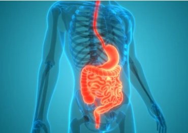
Digestive System
The digestive system develops as a simple blind-ended primitive gut tube, from which the digestive organs develop as outpouchings.
For ease of understanding, the development of the primitive gut tube can be summarized into the following categories which will each be discussed separately:
Development of the foregut
Development of the midgut
Development of the hindgut
Development of the Foregut
The structures that arise in the region of the foregut receive their blood supply from the celiac trunk (also known as celiac artery). The exceptions to this blood supply at the pharynx and the part of the respiratory system that is derived from the foregut.
The region of the foregut includes:
Pharynx
This is the most proximal region of the foregut which continues as the esophagus.
Esophagus
This structure is distinguishable by week four of development.
The lung bud develops as an endodermal outpouching of the esophagus, known as the respiratory diverticulum (refer to embryology of the respiratory system).
The esophageal epithelium and glands are derivatives of foregut endoderm, which esophageal muscles are derivatives of the surrounding mesoderm.
In the early stages of development, epithelial cells proliferate and fill the lumen of the esophagus, turning it into a solid rod of tissue. By week eight of development, this tube is recanalize and the esophagus becomes a hollow tube spanning from the pharynx to the stomach.
Stomach
This structure develops from a small dilation of the foregut endoderm.
The stomach has a dorsal border that is anchored to the posterior body wall by a double layer sheet of mesodermal tissue known as the dorsal mesogastrium. It also has a ventral border that is anchored to the ventral body wall by a double layer sheet of mesodermal tissue known as the ventral mesogastrium.
Rotation of the stomach:
A 90 degree clockwise rotation around the stomach’s longitudinal axis (or from a cranial perspective) brings it’s left side to a ventral position, and it’s right side to a dorsal position. This provides an embryologic explanation as to why the left vagus nerve innervates the ventral wall and the right vagus nerve innervates the dorsal wall. This rotation also gives rise to the greater and lesser curvatures of the stomach.
A rotation around is antero-posterior axis allows the pyloric region to move upwards while the cardiac region moves downwards, allowing the stomach to sit in its final position.
The rotation of the stomach brings the dorsal mesogastrium to the left and the ventral mesogastrium to the right, creating a small space posterior to the stomach known as the lesser peritoneal sac (or the omental bursa).
The dorsal mesogastrium enlarges to form the greater omentum, while the ventral mesogastrium forms the lesser omentum.
Proximal half of the duodenum
This structure develops into a C-shaped loop that sits on the right side of the abdominal cavity.
The rotation and growth of the stomach bring the duodenum to its final position towards the dorsal body wall. Through further development, this region loses its mesentery and becomes secondarily retroperitoneal.
The lumen of the primordial duodenum is filled with epithelium and only recanalizes in week six of development.
Liver and biliary apparatus
The liver, gall bladder, and biliary ductal system are derived from a hepatic diverticulum on the ventral mesogastrium (refer to stomach above) and extends into the septum transversum (refer to diaphragm).
The hepatic diverticulum gives rise to a larger region that develops into the liver, a smaller region that develops into the gall bladder, and the stalk connecting the diverticulum to the foregut will narrow to give rise to the bile duct.
Increased cellular proliferation on the right side of the liver results in a much larger right lobe, in comparison to the smaller left lobe of the liver.
The mesoderm surrounding the region of the surface of the liver will differentiate into the visceral peritoneum that overlies the liver.
The cranial region of the liver which develops while in contact with the septum transversum, remains uncovered by visceral peritoneum, providing an embryologic explanation for the region of the liver known as the bare area.
The ventral mesogastrium also gives rise to the falciform ligament which anchors the liver to the anterior body wall.
Pancreas
This structure is formed from the endodermal lining of the foregut that initially develop as two separate pancreatic buds that later unite.
The dorsal pancreatic duct forms as an outpouching of the dorsal wall of the duodenum, while the ventral pancreatic bud forms as an outpouching of the ventral wall of the duodenum. As the duodenum rotates during development, the two pancreatic buds are pulled together and unite.
The ventral pancreatic bud forms the uncinate process and inferior region of the head of the pancreas.
The dorsal pancreatic bud forms the superior region of the head, neck, and body of the pancreas.
As development progresses, the ductal system of both pancreatic buds will fuse to give rise to the main pancreatic duct.
Development of the Midgut
The structures that arise in the region of the midgut receive their blood supply from the superior mesenteric artery.
The region of the midgut includes:
Remaining parts of small intestine (distal half of duodenum, jejunum, and ileum)
By week five of development, this region begins elongating at a rate that is faster than the abdominal cavity is growing, resulting in the need for the formation of the primary intestinal loop:
The primary intestinal loop communicates with the yolk sac through the vitelline duct, while the superior mesenteric artery runs along the axis of this loop.
The cranial limb of this loop will develop into the distal half of the duodenum, the jejunum, and the proximal half of the ileum.
Parts of the large intestine (cecum, appendix, ascending colon, proximal two-thirds of the transverse colon)
The caudal limb of the primary intestinal loop (described above) will develop into the distal half of the ileum, cecum, ascending colon, and the proximal two-thirds of the transverse colon.
Between week six and eight of development, the primary intestinal loop will undergo the process of physiological herniation. The events occurring during this can be summarized as:
The primary intestinal loop will protrude into the umbilicus.
At the same time, the primary intestinal loop begins rotating 90 degrees in a counterclockwise direction around the axis of the superior mesentery artery. This rotation allows the cranial limb to move more caudally and to the right, and the caudal limb to move more cranially and to the left.
As the primary intestinal loop rotates, the jejunal and ileal regions of the small intestine continue to develop into intestinal loops while the cecum gives rise to the appendix.
By week ten of development, this developing loop of midgut structures will retract back into the abdominal cavity. At this stage, it rotates a further 180 degrees counterclockwise around the axis of the superior mesenteric artery (for a total of 270 degrees of counterclockwise rotation around the same axis). This final rotation brings the midgut structures into their final positions within the abdominal cavity.
During week eleven of development, the dorsal mesogastrium of the ascending colon will shorten and fold, and anchor it to the dorsal wall of the body, thereby making it secondarily retroperitoneal. Meanwhile, the remaining organs (distal half of duodenum, jejunum, ileum, cecum, and the transverse colon) are intraperitoneal as they are suspended by a short mesentery from the dorsal wall of the body.
Development of the Hindgut
The structures that arise in the region of the hindgut receive their blood supply from the inferior mesenteric artery.
The region of the hindgut includes:
Distal third of the transverse colon, descending colon, sigmoid colon, rectum, and the upper two-thirds of the anal canal
In the early stages of the development of the hindgut, it ends in a simple cavity known as the cloaca. This is an endoderm lined pouch that is lined by the cloacal membrane ventrally, and is composed of surface ectoderm.
By week seven of development, a wedge of mesoderm known as the urorectal septum will divide the cloaca into ventral and dorsal regions
The primitive urogenital sinus will develop ventrally. This will give rise to the bladder, urethra, vagina (in females), prostate gland and membranous urethra (in males).
The anorectal canal will develop dorsally. This will give rise to the rectum and upper anal canal.
As development progresses, the cloacal membrane will rupture to connect the developing superior two-thirds and inferior one-third of the anorectal canal. The junction between these two regions is marked by the pectinate line.
The superior two-thirds of the anorectal canal form the distal portion of the hindgut and is endoderm in origin.
The inferior one-third of the anorectal canal is derived from the surface ectoderm of the anal pit.
Peritoneal Cavity
Peritoneum is a serous membrane that forms the lining of the abdominal cavity, it supports the organs of the abdominal cavity and allows the passage of blood vessels, lymphatic vessels, and nerves.
The layers of peritoneum can be summarized as:
Parietal peritoneum - this lines the abdominal wall and receives somatic innervation, rendering it sensitive to localized pain.
Visceral peritoneum - this lines the viscera directly and receives autonomic innervation, rendering it sensitive to referred pain, or poorly localized sensations of discomfort.
Between the parietal and visceral layers of peritoneum, a potential space exists, known as the peritoneal cavity. The peritoneal cavity is filled with serous fluid.
Structures that are within the peritoneum and known as intraperitoneal structures while structures that are posterior to the peritoneum are know as retroperitoneal structures.
Examples of intraperitoneal organs include:
Abdominal esophagus
Stomach
Liver
Gall bladder
Spleen
Jejunum
Ileum
Cecum
Appendix
Sigmoid colon
Retroperitoneal organs may be primary or secondary in nature.
Primary retroperitoneal organs developed within, and as development progressed, stayed within the parietal peritoneum.
Examples of primary retroperitoneal organs include:
Esophagus
Rectum
Anus
Secondary retroperitoneal organs developed intraperitoneally, and as development progressed, their mesentery fused with the posterior abdominal wall, making them retroperitoneal in nature.
Examples of secondary retroperitoneal organs include:
Proximal half of duodenum
Pancreas
Ascending colon
Descending colon
Mesentery
Mesentery develops as a covering of mesenchyme that originates from the posterior abdominal wall and passes over the gut tube. As the primitive gut tube develops, initially it is in close contact with the posterior abdominal wall, in time, it begins pulling away from the posterior abdominal wall and towards the the region that will become the abdominal cavity. In doing so, the mesentery eventually develops into a structure that allows the suspension of the abdominal organs from the dorsal and ventral abdominal walls.
The dorsal mesentery will give rise to the mesenteries of the small and large intestines of the developed gastrointestinal tract, and will also give rise to the greater omentum. Examples of mesentery include:
Mesoduodenum - dorsal mesentery of the duodenum
Mesoappendix - dorsal mesentery of the appendix
Mesocolon - dorsal mesentery of the colon
Mesentery proper - dorsal mesentery of the jejunum and ileum
The ventral mesentery will give rise to the lesser omentum and the falciform ligament of the liver.
Both the greater omentum and the lesser omentum are composed of peritoneum that has folded over itself, creating what is described as a double layer of peritoneum.
Clinical Notes:
Foregut:
Duodenal atresia - this occurs if there is an incomplete recanalization of the duodenum, wherein some regions may remain occluded.
Midgut:
Meckel’s diverticulum - this occurs when a remnant of the vitelline duct persists, leading to pain and bleeding.
Congenital umbilical hernia - this occurs when the primary intestinal loop fails to retract back into the abdominal cavity as development progresses.
Hindgut:
Imperforate anus - this occurs when there is an anorectal malformation, resulting in an absent anal opening during birth.


